
Summit County’s premier center for nonsurgical orthopedics and regenerative medicine.

We deliver minimally invasive nonsurgical, advanced diagnostics, and personalized recovery plans that restore function and optimize performance. Through our specialized expertise, cutting-edge therapies, and individualized care, we ensure our patients return to the activities they love, stronger and more resilient than before.

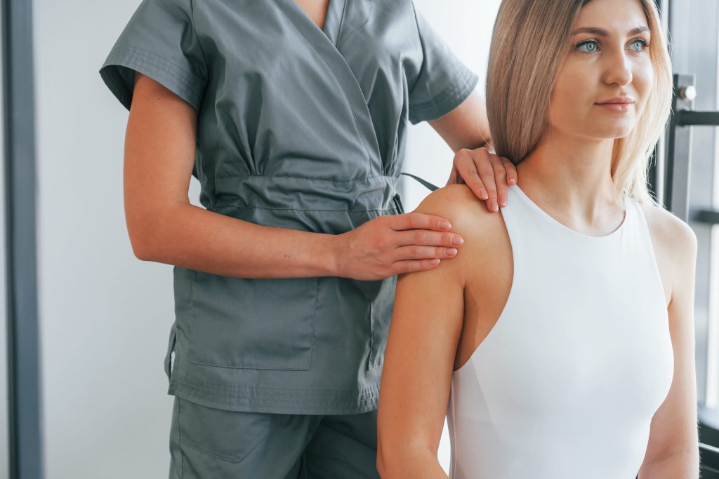
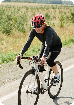


Our independent, locally owned clinic specializes in advanced, nonsurgical treatments designed to repair tissue and restore function.

Non-surgical treatment options address joint arthritis, labral injuries, rotator cuff injuries, and occasionally referral pain from the neck.
Injuries range from rotator cuff tendinopathy, tendon tears, and subacromial tendon impingement.
Injury to the ligaments connecting the collarbone and the shoulder blade.
Compression of the rotator cuff tendons as they pass under the acromion (component of the scapular bone), often due to suboptimal movement of the scapula.
Inflammation of the bursa, a small fluid-filled sac, that cushions the rotator cuff tendons.
Inflammation and thickening of the joint capsule causing shoulder pain, stiffness and loss of range of motion.
"Wear-and-tear" of the articular cartilage of the AC joint or glenohumeral joint (main shoulder joint). Shoulder joint arthritis presents as pain with active shoulder movement. In more advanced stages, shoulder OA can limit range of motion of the shoulder, particularly with overhead movements and reaching behind the back.
Anterior shoulder pain due to biceps tendinitis (inflammation), tendinopathy (injured tendon fibers), or a tendon tear.
Injury to the fibrous cartilage lining the glenoid (shoulder socket). Injuries range from degeneration (wear-and-tear), to posterior tears, to SLAP tears and bankart lesions (from a shoulder dislocation). Depending on the type of injury, treatment options may be optimized non-surgically, while many require a surgical repair.

Common causes of hip pain include osteoarthritis, labral injury, or surrounding tendon injuries. These conditions are often treated non-surgically.
Degenerative arthritis of the ball-and-socket hip joint. Pain often refers to the groin and top of the thigh.
The labrum is a dense fibrocartilage tissue lining the acetabulum (hip socket) that acts to deepen the socket, stabilize the hip, absorb impact activities, and create a synovial fluid seal for smooth movement and to provide nutrients to the joint. Injuries should be diagnosed and characterized with advanced imaging (MRI).
Inflammation of the bursa on the outside of the hip (greater trochanter), or more commonly, disorganization and damage (tendinopathy) of the hip stabilizing tendons that attach to the greater trochanter.
Inflammation (tendinitis) or cellular disorganization and damage (tendinopathy) of the tendons around the hip. Affected tendons may include the quadriceps, iliopsoas, sartorius, or gluteus medius and minimus.
Sensation of a snapping sensation, often with associated pain during movement. "External snapping hip" involves the IT band, while "internal snapping hip" involves the iliopsoas or labrum. Risk factors include repetitive hip motions (running, dance, soccer), tight hip muscles/tendons (iliopsoas, IT band), inadequate warm-ups, flat feet, certain anatomical variations (like a prominent greater trochanter or leg length difference), trauma, and sometimes prior hip/knee surgery or arthritis.
Symptoms are often characterized as sharp to dull pain in the low back and gluteal regions, worse with prolonged positions and positional changes.
Irritation or spasm of the piriformis muscle that commonly causes pain in the buttock, hip and upper leg.Occasionally, this condition compresses the sciatic nerve resulting in referral pain down the leg.

The knee is highly susceptible to wear and tear and soft tissue injuries.
"Wear-and-tear" of the articular cartilage in the knee. There are three articular compartments within the knee--the medial and lateral tibiofemoral compartments and the patellofemoral compartment. Degenerative injuries may occur to one, or all, of the compartments. Symptoms include localized pain, stiffness and swelling.
The meniscus is a crescent-shape dense fibrocartilage that acts as a shock absorber for the knee. There are two menisci, one on the medial (inside) side and one at the lateral (outside) side of the joint where the femur (thigh) and tibia (lower leg) bones articulate. Meniscus injuries can range from small stable tears to complex or degenerative tears. Symptoms may include localized knee pain (at the inside or outside of the joint), swelling and catching.
Pain involving the knee cap. This condition is often due to the mechanical alignment of the patellofemoral joint, and often responds well to physical therapy and bracing.
Inflammation (tendinitis) or cellular disorganization and damage (tendinopathy) of the tendons around the knee. Symptoms often present as anterior knee pain made worse with activities such as running and jumping.
Ligaments help to stabilize the knee, both internally (ACL and PCL) and externally (MCL and LCL). Injuries can be isolated to a single ligament, or affect multiple ligaments at once. Ligament injuries range from grade I (mild) to grade III (severe). Depending on the severity, ligament injuries are often treated non-surgically, though some injuries require a surgical repair.
Bursa are small fluid-filled sacs that reduce friction between layers of soft tissue (such as tendons, muscle, and skin) and between soft tissue and bone. Bursitis occurs when these sacs become inflamed causing swelling and localized pain. Common affected sites include the prepatellar bursa (over the top of the knee), the infrapatellar bursa (blow the knee cap), and the Pes Anserine bursa (on the inner side of the knee).
The IT band (ITB) is a thick band of tissue running from your hip down the outside of your thigh to your knee. The ITB can become irritated or injured at the hip or further down at the knee, and is often caused by an overuse injury from activities such as cycling or running.
Pain over the top and inside of the lower leg due to a stress reaction involving the tibial bone (shin bone). This overuse injury is due to repetitive strain of the calf muscles (tibialis posterior and soleus) and their attachment to the tibial bone. Pain symptoms commonly occur at the beginning and/or end of a high impact activity, such as sprinting, distance running, soccer and dancing.
Compression of the common fibular nerve near the knee (most commonly as it crosses the fibular head). Symptoms may include pain, numbness and tingling that refers down the outside and front of the lower leg to the top of the foot. More severe injuries include foot and ankle weakness.

Common non-surgical issues involve nerve compression, tendon inflammation (tendinitis), tendon degeneration (tendinopathy), tendon tears, repetitive strain injuries, and joint arthritis.
Compression of the median nerve as it passes through the carpal tunnel in the wrist.
Inflammation of the tendons on the thumb side of the wrist, causing pain with gripping.
Inflammation and thickening of the tendon sheath and pulley (ligament that passes over the tendon) causing the finger to catch and lock.
Tendon inflammation (tendinitis) or tendon injury (tendinopathy or tear) involving any of the tendons in the hand and wrist.
Fluid-filled, painful lumps (non-cancerous) caused by inflammation. These cysts often occur in the back of the wrist or hand.
Degenerative joint disease (wear-and-tear), especially common at the base of the thumb.
clicking and pain over the wrist due to instability (subluxation) and inflammation of the extensor carpi ulnaris (ECU) tendon secondary to overuse and tendon sheath injury.
An overuse tendinitis (inflammation) resulting in pain, and occasionally swelling, at the back of the forearm where tendons of the thumb (first compartment) cross over extensor tendons of the wrist (second compartment).
Compression of the ulnar nerve at the wrist as the ulnar nerve courses through Guyon's Canal (often due to a ganglion cyst or pressure from handlebars while cycling). Depending on the involved nerve fibers, symptoms range from medial (side of the pinky) hand numbness to hand weakness, or both.
An injury at the base of the thumb involving the ulnar collateral ligament (UCL). The most commonly injured ligament in the thumb, this injury presents as pain and weakness in thumb pinch and grasp.
Injury to the cartilage and ligaments that stabilize the pinky-finger side (ulnar-sided) of the wrist. Symptoms often include ulnar-sided wrist pain, swelling and weakness.

Common conditions involve tendon injuries near the ankle joint, injuries to ligaments that support ankle, joint injuries and degenerative conditions, injuries to the plantar fascia, and nerve irritation and impingement.
Thickening, loss of normal cellular architecture and tearing of the band of tissue (plantar fascia) that runs across the bottom of the foot. Symptoms are often sharp pain at the heel of the foot, usually worse in the morning, and with prolonged standing and walking.
Inflammation (tendinitis) or chronic degeneration and micro-tears (tendinopathy) of the Achilles tendon. The injury can occur at the mid-portion of the heal cord (non-insertional) or at the insertion with the heel (calcaneus bone). Symptoms are often associated with running, hiking, and basketball.
Ligaments help support the ankle joint, and are commonly injured during an "ankle roll" during activities such as running, hiking or volleyball. These injuries often occur when rolling the foot to the inside (inversion injury), affecting the ATFL (anterior tibiofibular ligament) at the top of the ankle. Symptoms include localized pain and swelling, while more severe injuries may have associated bruising and ankle instability.
Compression of the posterior tibial nerve at the medial (inner side) ankle. Symptoms may include pain, numbness and tingling at the inner ankle that refers to the heel and sole of the foot.
Thickening of the tissue around a digital nerve between the toes causing localized pain between the affected toes, and often a sensation of walking on a pebble. Symptoms are often worse with tight footwear and stiff boots (ie, ski boots).
Degenerative arthritis in the joints of the ankle or foot. Symptoms are often worse with standing and impact activities such as walking and running.
Bony overgrowth of the metatarsal phalangeal joint (MTP) of the great toe causing the toe to point inward toward the other toes. This condition causes pain, redness, and difficulty wearing tight shoes or boots.
Inflammation (tendinitis) or chronic degeneration and micro-tears (tendinopathy) of tendons surrounding the ankle. Affected tendons may be at the medial (inside) or lateral (outside) side of the ankle. Symptoms include localized pain at the ankle over the affected tendon, which is often worse with walking (particularly on uneven surfaces) and high intensity activities.

Non-surgical management is the primary approach for most cases of low back pain, which often stems from muscle, disc, or joint issues.
Injury to the muscles or ligaments of the lower back.
Nerve compression in the low back that causes referral pain and weakness into the affected leg.
Injury to the intervertebral disc involving the annular fibers and/or central nucleus that can compress an existing nerve (radiculopathy) causing pain and weakness into the leg.
Narrowing of the spinal canal, often the result of age-related degenerative disc disease, a disc herniation, spondylolisthesis (vertebral body slips out of alignment). Symptoms may include generalized back back pain and weakness worse with walking, that refers to the thighs or lower legs.
Arthritis/inflammation of the small joints along the back of the spine. Typically presents as generalized, or localized, low back and buttock pain worse with standing.
Stress fracture of the lumbar spine that is common in young athletes, particularly those who repeatedly bend backwards (such as gymnastics). Commonly presents as localized pain in the low back.
Pain involving an intervertebral disc that is commonly caused by a tear in the disc's annular fibers. Symptoms are often characterized by a deep ache/pain that is worse with bending, prolonged sitting or standing, and lifting.
Irritation or spasm of the piriformis muscle that commonly causes pain in the buttock, hip and upper leg. Though less common, this condition can compresses the sciatic nerve resulting in referral pain down the leg
"Tailbone pain" that is commonly caused by injuring a joint in the coccyx (spinal segment at the bottom of your spine) by a traumatic event, such as a fall. The symptoms are characterized by a sharp to dull pain at the tailbone that is worse with sitting, and transitioning from sitting to standing.
Age-related changes where the intervertebral discs lose hydration and height. Associated conditions include discogenic back pain, facet pain/arthritis, central canal stenosis, and nerve impingement (radiculitis).

Common conditions include tendon injuries near the elbow, elbow ligament injury, elbow joint degenerative arthritis, elbow bursitis, and nerve irritation and impingement (in the upper arm, elbow, or forearm)
Triceps tendinopathy (cellular disorganization of the tendon) or a tendon tear that most frequently occurs where the tendon attaches to the olecranon (back of the elbow). Pain symptoms are localized to the back of the arm and elbow, and are often worse with lifting, throwing and resisted triceps extension exercises.
The UCL is located at the inside of the elbow, and its primary function is to stabilize the elbow. UCL injuries range from stretching to tearing of the ligaments, causing pain and instability at the inner side of the elbow. Injuries are common among throwing athletes.
Pain at the anterior shoulder (proximal biceps tendon) or anterior elbow (insertional biceps tendon) due to tendinitis (inflammation), tendinopathy (tendon disorganization and micro-tears), or a tendon tear. Proximal long head biceps tendinitis may be associated with a labral injury, while a complete tear of this tendon is relatively painless and with an associated "popeye sign" due to the muscle loosening and balliing up above the front of the elbow.
Pain at the medial (inside of elbow) or lateral (outside of elbow) elbow and forearm due to tendinitis (inflammation), tendinopathy (chronic tendon disorganization and micro-tears), or a tendon tear. Pain is worse with repetitive activities, or even with simple actions like holding a mug of coffee.
Pain and swelling at the back of elbow. The olecranon bursa (small fluid-filled sac) provides cushion between the triceps tendon and olecranon (back of elbow) bone. Pain and swelling occurs with the bursa becomes inflamed and swollen. Symptoms are often worse with direct pressure over the elbow.
Degenerative cartilage breakdown in the elbow joint. Symptoms include pain with active range of motion, and loss of elbow extension (straightening).
The second most common neuropathy, characterized by compression or "snapping" of the ulnar nerve as it passes through the cubital tunnel at the inside of the elbow. Symptoms include medial elbow pain and numbness and tingling to the inside of the hand and commonly includes weakness of the ulnar-innervated hand muscles resulting in a loss of dexterity.
Compression of the median nerve as it courses through the palmar aspect of the wrist. Symptoms include numbness and tingling with associated pain involving the palm, thumb, index and middles fingers. Occasionally symptoms involve the entire hand. Severe cases include grip strength weakness and muscle atrophy (at the base of the thumb). First-line treatments include bracing and PT. Interventional options include steroid or platelet-rich plasma (PRP) injections. Recalcitrant cases are treated with a carpal tunnel release surgery.
Median nerve compression at the front of the elbow and forearm, typically as it passes through the pronator teres muscle. This causes a deep pain at the front of elbow and forearm with numbness, tingling and weakness in the forearm and hand. Symptoms are often worse with repetitive gripping and forearm pronation (turning the wrist toward the floor). Unlike carpal tunnel syndrome, sensation is spared over the pad of the thumb (thenar eminence).
Compression of the deep motor branch of the radial nerve as it passes through the radial tunnel in the back side of the forearm. This causes weakness in the extensor muscles of the thumb and hand with no associated sensory loss or numbness and tingling.
A relatively uncommon condition caused by the compression of the posterior interosseous nerve (a branch of the radial nerve) near its origin in the spiral groove (back of elbow/forearm). Unlike PIN syndrome, symptoms are often limited to isolated pain and tenderness at the back of the forearm. Symptoms are often worse with wrist extension and forearm supination (rotating the wrist upward). First-line treatments are PT and activity modifications. Interventional options include nerve hydrodissection. For recalcitrant cases, a surgical release is indicated.

Common non-surgical conditions that cause neck pain, stiffness, or referral pain to the arms.
Degenerative changes involving intervertebral discs and facet joints. Associated symptoms include generalized or local neck pain.
Localized or regional neck pain caused by facet arthritis or injury to the facet capsule (often the result of trauma).
Facet capsular injury, or muscle or ligament injury as a result of trauma.
Injury to the intervertebral disc involving the annular fibers and/or central nucleus, potentially pressing on nerves. Symptoms range from neck pain to pain that travels down the arm.
Nerve compression in the neck that causes referral pain and weakness into the affected arm.
Muscle spasms and trigger points causing stiffness and neck pain that may refer to the shoulders and arms.
Referral pain to the back of the head due to occipital nerve irritation (occipital neuralgia) or symptomatic facet joints.
Commonly Treated Nerve Conditions:
The most common compressive neuropathy. The median nerve is compressed as it courses through the palmar aspect of the wrist. Symptoms include numbness and tingling with associated pain involving the palm, thumb, index and middle fingers. Patients often experience weakness in grip strength. Occasionally symptoms involve the entire hand. Severe cases involve muscle atrophy at the base of the thumb.
Compression of the deep motor branch of the radial nerve as it passes through the radial tunnel in the back side of the forearm. This causes weakness in the extensor muscles of the thumb and hand with no associated sensory loss or numbness and tingling.
The second most common neuropathy, characterized by compression or "snapping" of the ulnar nerve as it passes through the cubital tunnel at the inside of the elbow. Symptoms include medial elbow pain and numbness and tingling to the inside of the hand and commonly includes weakness of the ulnar-innervated hand muscles resulting in a loss of dexterity.
Median nerve compression at the front of the elbow and forearm, typically as it passes through the pronator teres muscle. This causes a deep pain at the front of elbow and forearm with numbness, tingling and weakness in the forearm and hand. Symptoms are often worse with repetitive gripping and forearm pronation (turning the wrist toward the floor). Unlike carpal tunnel syndrome, sensation is spared over the pad of the thumb (thenar eminence).
Compression of the ulnar nerve at the wrist as the ulnar nerve courses through Guyon's Canal (often due to a ganglion cyst or pressure from handlebars while cycling). Depending on the involved nerve fibers, symptoms range from medial (side of the pinky) hand numbness to hand weakness, or both.
Compression of the posterior tibial nerve at the medial (inner side) ankle. Symptoms may include pain, numbness and tingling at the inner ankle that refers to the heel and sole of the foot.
Compression of the common fibular nerve near the knee (most commonly as it crosses the fibular head). Symptoms may include pain, numbness and tingling that refers down the outside and front of the lower leg to the top of the foot. More severe injuries include foot and ankle weakness.
Thickening of the tissue around a digital nerve between the toes causing localized pain between the affected toes, and often a sensation of walking on a pebble. Symptoms are often worse with tight footwear and stiff boots (ie, ski boots).
Nerve compression in the neck that causes referral pain and weakness into the affected arm.
Nerve compression in the low back that causes referral pain and weakness into the affected leg.

Non-surgical treatment options address joint arthritis, labral injuries, rotator cuff injuries, and occasionally referral pain from the neck.
Injuries range from rotator cuff tendinopathy, tendon tears, and subacromial tendon impingement.
Injury to the ligaments connecting the collarbone and the shoulder blade.
Compression of the rotator cuff tendons as they pass under the acromion (component of the scapular bone), often due to suboptimal movement of the scapula.
Inflammation of the bursa, a small fluid-filled sac, that cushions the rotator cuff tendons.
Inflammation and thickening of the joint capsule causing shoulder pain, stiffness and loss of range of motion.
"Wear-and-tear" of the articular cartilage of the AC joint or glenohumeral joint (main shoulder joint). Shoulder joint arthritis presents as pain with active shoulder movement. In more advanced stages, shoulder OA can limit range of motion of the shoulder, particularly with overhead movements and reaching behind the back.
Anterior shoulder pain due to biceps tendinitis (inflammation), tendinopathy (injured tendon fibers), or a tendon tear.
Injury to the fibrous cartilage lining the glenoid (shoulder socket). Injuries range from degeneration (wear-and-tear), to posterior tears, to SLAP tears and bankart lesions (from a shoulder dislocation). Depending on the type of injury, treatment options may be optimized non-surgically, while many require a surgical repair.
Common causes of hip pain include osteoarthritis, labral injury, or surrounding tendon injuries. These conditions are often treated non-surgically.
Degenerative arthritis of the ball-and-socket hip joint. Pain often refers to the groin and top of the thigh.
The labrum is a dense fibrocartilage tissue lining the acetabulum (hip socket) that acts to deepen the socket, stabilize the hip, absorb impact activities, and create a synovial fluid seal for smooth movement and to provide nutrients to the joint. Injuries should be diagnosed and characterized with advanced imaging (MRI).
Inflammation of the bursa on the outside of the hip (greater trochanter), or more commonly, disorganization and damage (tendinopathy) of the hip stabilizing tendons that attach to the greater trochanter.
Inflammation (tendinitis) or cellular disorganization and damage (tendinopathy) of the tendons around the hip. Affected tendons may include the quadriceps, iliopsoas, sartorius, or gluteus medius and minimus.
Sensation of a snapping sensation, often with associated pain during movement. "External snapping hip" involves the IT band, while "internal snapping hip" involves the iliopsoas or labrum. Risk factors include repetitive hip motions (running, dance, soccer), tight hip muscles/tendons (iliopsoas, IT band), inadequate warm-ups, flat feet, certain anatomical variations (like a prominent greater trochanter or leg length difference), trauma, and sometimes prior hip/knee surgery or arthritis.
Symptoms are often characterized as sharp to dull pain in the low back and gluteal regions, worse with prolonged positions and positional changes.
Irritation or spasm of the piriformis muscle that commonly causes pain in the buttock, hip and upper leg.Occasionally, this condition compresses the sciatic nerve resulting in referral pain down the leg.
The knee is highly susceptible to wear and tear and soft tissue injuries.
"Wear-and-tear" of the articular cartilage in the knee. There are three articular compartments within the knee--the medial and lateral tibiofemoral compartments and the patellofemoral compartment. Degenerative injuries may occur to one, or all, of the compartments. Symptoms include localized pain, stiffness and swelling.
The meniscus is a crescent-shape dense fibrocartilage that acts as a shock absorber for the knee. There are two menisci, one on the medial (inside) side and one at the lateral (outside) side of the joint where the femur (thigh) and tibia (lower leg) bones articulate. Meniscus injuries can range from small stable tears to complex or degenerative tears. Symptoms may include localized knee pain (at the inside or outside of the joint), swelling and catching.
Pain involving the knee cap. This condition is often due to the mechanical alignment of the patellofemoral joint, and often responds well to physical therapy and bracing.
Inflammation (tendinitis) or cellular disorganization and damage (tendinopathy) of the tendons around the knee. Symptoms often present as anterior knee pain made worse with activities such as running and jumping.
Ligaments help to stabilize the knee, both internally (ACL and PCL) and externally (MCL and LCL). Injuries can be isolated to a single ligament, or affect multiple ligaments at once. Ligament injuries range from grade I (mild) to grade III (severe). Depending on the severity, ligament injuries are often treated non-surgically, though some injuries require a surgical repair.
Bursa are small fluid-filled sacs that reduce friction between layers of soft tissue (such as tendons, muscle, and skin) and between soft tissue and bone. Bursitis occurs when these sacs become inflamed causing swelling and localized pain. Common affected sites include the prepatellar bursa (over the top of the knee), the infrapatellar bursa (blow the knee cap), and the Pes Anserine bursa (on the inner side of the knee).
The IT band (ITB) is a thick band of tissue running from your hip down the outside of your thigh to your knee. The ITB can become irritated or injured at the hip or further down at the knee, and is often caused by an overuse injury from activities such as cycling or running.
Pain over the top and inside of the lower leg due to a stress reaction involving the tibial bone (shin bone). This overuse injury is due to repetitive strain of the calf muscles (tibialis posterior and soleus) and their attachment to the tibial bone. Pain symptoms commonly occur at the beginning and/or end of a high impact activity, such as sprinting, distance running, soccer and dancing.
Compression of the common fibular nerve near the knee (most commonly as it crosses the fibular head). Symptoms may include pain, numbness and tingling that refers down the outside and front of the lower leg to the top of the foot. More severe injuries include foot and ankle weakness.
Common non-surgical issues involve nerve compression, tendon inflammation (tendinitis), tendon degeneration (tendinopathy), tendon tears, repetitive strain injuries, and joint arthritis.
Compression of the median nerve as it passes through the carpal tunnel in the wrist.
Inflammation of the tendons on the thumb side of the wrist, causing pain with gripping.
Inflammation and thickening of the tendon sheath and pulley (ligament that passes over the tendon) causing the finger to catch and lock.
Tendon inflammation (tendinitis) or tendon injury (tendinopathy or tear) involving any of the tendons in the hand and wrist.
Fluid-filled, painful lumps (non-cancerous) caused by inflammation. These cysts often occur in the back of the wrist or hand.
Degenerative joint disease (wear-and-tear), especially common at the base of the thumb.
clicking and pain over the wrist due to instability (subluxation) and inflammation of the extensor carpi ulnaris (ECU) tendon secondary to overuse and tendon sheath injury.
An overuse tendinitis (inflammation) resulting in pain, and occasionally swelling, at the back of the forearm where tendons of the thumb (first compartment) cross over extensor tendons of the wrist (second compartment).
Compression of the ulnar nerve at the wrist as the ulnar nerve courses through Guyon's Canal (often due to a ganglion cyst or pressure from handlebars while cycling). Depending on the involved nerve fibers, symptoms range from medial (side of the pinky) hand numbness to hand weakness, or both.
An injury at the base of the thumb involving the ulnar collateral ligament (UCL). The most commonly injured ligament in the thumb, this injury presents as pain and weakness in thumb pinch and grasp.
Injury to the cartilage and ligaments that stabilize the pinky-finger side (ulnar-sided) of the wrist. Symptoms often include ulnar-sided wrist pain, swelling and weakness.
Common conditions involve tendon injuries near the ankle joint, injuries to ligaments that support ankle, joint injuries and degenerative conditions, injuries to the plantar fascia, and nerve irritation and impingement.
Thickening, loss of normal cellular architecture and tearing of the band of tissue (plantar fascia) that runs across the bottom of the foot. Symptoms are often sharp pain at the heel of the foot, usually worse in the morning, and with prolonged standing and walking.
Inflammation (tendinitis) or chronic degeneration and micro-tears (tendinopathy) of the Achilles tendon. The injury can occur at the mid-portion of the heal cord (non-insertional) or at the insertion with the heel (calcaneus bone). Symptoms are often associated with running, hiking, and basketball.
Ligaments help support the ankle joint, and are commonly injured during an "ankle roll" during activities such as running, hiking or volleyball. These injuries often occur when rolling the foot to the inside (inversion injury), affecting the ATFL (anterior tibiofibular ligament) at the top of the ankle. Symptoms include localized pain and swelling, while more severe injuries may have associated bruising and ankle instability.
Compression of the posterior tibial nerve at the medial (inner side) ankle. Symptoms may include pain, numbness and tingling at the inner ankle that refers to the heel and sole of the foot.
Thickening of the tissue around a digital nerve between the toes causing localized pain between the affected toes, and often a sensation of walking on a pebble. Symptoms are often worse with tight footwear and stiff boots (ie, ski boots).
Degenerative arthritis in the joints of the ankle or foot. Symptoms are often worse with standing and impact activities such as walking and running.
Bony overgrowth of the metatarsal phalangeal joint (MTP) of the great toe causing the toe to point inward toward the other toes. This condition causes pain, redness, and difficulty wearing tight shoes or boots.
Inflammation (tendinitis) or chronic degeneration and micro-tears (tendinopathy) of tendons surrounding the ankle. Affected tendons may be at the medial (inside) or lateral (outside) side of the ankle. Symptoms include localized pain at the ankle over the affected tendon, which is often worse with walking (particularly on uneven surfaces) and high intensity activities.
Non-surgical management is the primary approach for most cases of low back pain, which often stems from muscle, disc, or joint issues.
Injury to the muscles or ligaments of the lower back.
Nerve compression in the low back that causes referral pain and weakness into the affected leg.
Injury to the intervertebral disc involving the annular fibers and/or central nucleus that can compress an existing nerve (radiculopathy) causing pain and weakness into the leg.
Narrowing of the spinal canal, often the result of age-related degenerative disc disease, a disc herniation, spondylolisthesis (vertebral body slips out of alignment). Symptoms may include generalized back back pain and weakness worse with walking, that refers to the thighs or lower legs.
Arthritis/inflammation of the small joints along the back of the spine. Typically presents as generalized, or localized, low back and buttock pain worse with standing.
Stress fracture of the lumbar spine that is common in young athletes, particularly those who repeatedly bend backwards (such as gymnastics). Commonly presents as localized pain in the low back.
Pain involving an intervertebral disc that is commonly caused by a tear in the disc's annular fibers. Symptoms are often characterized by a deep ache/pain that is worse with bending, prolonged sitting or standing, and lifting.
Irritation or spasm of the piriformis muscle that commonly causes pain in the buttock, hip and upper leg. Though less common, this condition can compresses the sciatic nerve resulting in referral pain down the leg
"Tailbone pain" that is commonly caused by injuring a joint in the coccyx (spinal segment at the bottom of your spine) by a traumatic event, such as a fall. The symptoms are characterized by a sharp to dull pain at the tailbone that is worse with sitting, and transitioning from sitting to standing.
Age-related changes where the intervertebral discs lose hydration and height. Associated conditions include discogenic back pain, facet pain/arthritis, central canal stenosis, and nerve impingement (radiculitis).
Common conditions include tendon injuries near the elbow, elbow ligament injury, elbow joint degenerative arthritis, elbow bursitis, and nerve irritation and impingement (in the upper arm, elbow, or forearm)
Triceps tendinopathy (cellular disorganization of the tendon) or a tendon tear that most frequently occurs where the tendon attaches to the olecranon (back of the elbow). Pain symptoms are localized to the back of the arm and elbow, and are often worse with lifting, throwing and resisted triceps extension exercises.
The UCL is located at the inside of the elbow, and its primary function is to stabilize the elbow. UCL injuries range from stretching to tearing of the ligaments, causing pain and instability at the inner side of the elbow. Injuries are common among throwing athletes.
Pain at the anterior shoulder (proximal biceps tendon) or anterior elbow (insertional biceps tendon) due to tendinitis (inflammation), tendinopathy (tendon disorganization and micro-tears), or a tendon tear. Proximal long head biceps tendinitis may be associated with a labral injury, while a complete tear of this tendon is relatively painless and with an associated "popeye sign" due to the muscle loosening and balliing up above the front of the elbow.
Pain at the medial (inside of elbow) or lateral (outside of elbow) elbow and forearm due to tendinitis (inflammation), tendinopathy (chronic tendon disorganization and micro-tears), or a tendon tear. Pain is worse with repetitive activities, or even with simple actions like holding a mug of coffee.
Pain and swelling at the back of elbow. The olecranon bursa (small fluid-filled sac) provides cushion between the triceps tendon and olecranon (back of elbow) bone. Pain and swelling occurs with the bursa becomes inflamed and swollen. Symptoms are often worse with direct pressure over the elbow.
Degenerative cartilage breakdown in the elbow joint. Symptoms include pain with active range of motion, and loss of elbow extension (straightening).
The second most common neuropathy, characterized by compression or "snapping" of the ulnar nerve as it passes through the cubital tunnel at the inside of the elbow. Symptoms include medial elbow pain and numbness and tingling to the inside of the hand and commonly includes weakness of the ulnar-innervated hand muscles resulting in a loss of dexterity.
Compression of the median nerve as it courses through the palmar aspect of the wrist. Symptoms include numbness and tingling with associated pain involving the palm, thumb, index and middles fingers. Occasionally symptoms involve the entire hand. Severe cases include grip strength weakness and muscle atrophy (at the base of the thumb). First-line treatments include bracing and PT. Interventional options include steroid or platelet-rich plasma (PRP) injections. Recalcitrant cases are treated with a carpal tunnel release surgery.
Median nerve compression at the front of the elbow and forearm, typically as it passes through the pronator teres muscle. This causes a deep pain at the front of elbow and forearm with numbness, tingling and weakness in the forearm and hand. Symptoms are often worse with repetitive gripping and forearm pronation (turning the wrist toward the floor). Unlike carpal tunnel syndrome, sensation is spared over the pad of the thumb (thenar eminence).
Compression of the deep motor branch of the radial nerve as it passes through the radial tunnel in the back side of the forearm. This causes weakness in the extensor muscles of the thumb and hand with no associated sensory loss or numbness and tingling.
A relatively uncommon condition caused by the compression of the posterior interosseous nerve (a branch of the radial nerve) near its origin in the spiral groove (back of elbow/forearm). Unlike PIN syndrome, symptoms are often limited to isolated pain and tenderness at the back of the forearm. Symptoms are often worse with wrist extension and forearm supination (rotating the wrist upward). First-line treatments are PT and activity modifications. Interventional options include nerve hydrodissection. For recalcitrant cases, a surgical release is indicated.
Common non-surgical conditions that cause neck pain, stiffness, or referral pain to the arms.
Degenerative changes involving intervertebral discs and facet joints. Associated symptoms include generalized or local neck pain.
Localized or regional neck pain caused by facet arthritis or injury to the facet capsule (often the result of trauma).
Facet capsular injury, or muscle or ligament injury as a result of trauma.
Injury to the intervertebral disc involving the annular fibers and/or central nucleus, potentially pressing on nerves. Symptoms range from neck pain to pain that travels down the arm.
Nerve compression in the neck that causes referral pain and weakness into the affected arm.
Muscle spasms and trigger points causing stiffness and neck pain that may refer to the shoulders and arms.
Referral pain to the back of the head due to occipital nerve irritation (occipital neuralgia) or symptomatic facet joints.
Commonly Treated Nerve Conditions:
The most common compressive neuropathy. The median nerve is compressed as it courses through the palmar aspect of the wrist. Symptoms include numbness and tingling with associated pain involving the palm, thumb, index and middle fingers. Patients often experience weakness in grip strength. Occasionally symptoms involve the entire hand. Severe cases involve muscle atrophy at the base of the thumb.
Compression of the deep motor branch of the radial nerve as it passes through the radial tunnel in the back side of the forearm. This causes weakness in the extensor muscles of the thumb and hand with no associated sensory loss or numbness and tingling.
The second most common neuropathy, characterized by compression or "snapping" of the ulnar nerve as it passes through the cubital tunnel at the inside of the elbow. Symptoms include medial elbow pain and numbness and tingling to the inside of the hand and commonly includes weakness of the ulnar-innervated hand muscles resulting in a loss of dexterity.
Median nerve compression at the front of the elbow and forearm, typically as it passes through the pronator teres muscle. This causes a deep pain at the front of elbow and forearm with numbness, tingling and weakness in the forearm and hand. Symptoms are often worse with repetitive gripping and forearm pronation (turning the wrist toward the floor). Unlike carpal tunnel syndrome, sensation is spared over the pad of the thumb (thenar eminence).
Compression of the ulnar nerve at the wrist as the ulnar nerve courses through Guyon's Canal (often due to a ganglion cyst or pressure from handlebars while cycling). Depending on the involved nerve fibers, symptoms range from medial (side of the pinky) hand numbness to hand weakness, or both.
Compression of the posterior tibial nerve at the medial (inner side) ankle. Symptoms may include pain, numbness and tingling at the inner ankle that refers to the heel and sole of the foot.
Compression of the common fibular nerve near the knee (most commonly as it crosses the fibular head). Symptoms may include pain, numbness and tingling that refers down the outside and front of the lower leg to the top of the foot. More severe injuries include foot and ankle weakness.
Thickening of the tissue around a digital nerve between the toes causing localized pain between the affected toes, and often a sensation of walking on a pebble. Symptoms are often worse with tight footwear and stiff boots (ie, ski boots).
Nerve compression in the neck that causes referral pain and weakness into the affected arm.
Nerve compression in the low back that causes referral pain and weakness into the affected leg.
Schedule a consultation to explore non-surgical treatment options that align with your active lifestyle.